
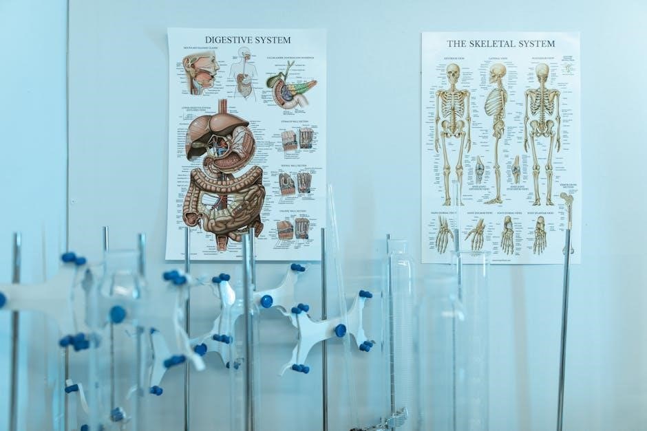
The skeletal system, comprising 206 bones, provides structural support, protects organs, facilitates movement, produces blood cells, and stores minerals, forming the body’s framework essential for human function and movement.
1.1 Overview of the Skeletal System
The skeletal system is the body’s framework, consisting of 206 bones, cartilage, tendons, and ligaments. It provides structural support, protects internal organs, and facilitates movement through joints. The adult skeleton is divided into the axial (central) and appendicular (limbs and pelvis) systems. Bones are made of calcium and phosphorus, giving them strength and durability. This system is dynamic, constantly remodeling to maintain health and function throughout life;
1.2 Importance of the Skeletal System in the Human Body
The skeletal system is vital for overall health, providing structural support and protection for organs like the brain and heart. It facilitates movement through joints and muscles, enabling mobility. Additionally, it produces blood cells in bone marrow and stores minerals such as calcium and phosphorus, essential for bodily functions. This system also plays a role in body shape and stability, making it indispensable for human survival and functionality.

Functions of the Skeletal System

The skeletal system performs essential functions, including providing structural support, protecting vital organs, facilitating movement, producing blood cells, and storing minerals for overall bodily function.
2.1 Support and Shape to the Body
The skeletal system provides structural support and maintains the body’s shape by forming a framework for tissues and organs. Bones act as attachment points for muscles, enabling posture and movement. The axial skeleton, including the skull, spine, ribs, and sternum, establishes the body’s central structure, while the appendicular skeleton, comprising limbs and girdles, extends this framework. Without the skeleton, the body would lack form and stability, making it unable to function effectively.
2.2 Protection of Vital Organs
The skeletal system protects vital organs by surrounding them with bony structures. The skull encases the brain, while the ribcage safeguards the heart, lungs, and liver. Vertebrae shield the spinal cord, and the pelvis protects reproductive organs. Bones act as a protective barrier, absorbing impacts and reducing damage to internal organs, ensuring the body’s essential systems remain safe and functional. This protective function is crucial for overall health and survival.
2.3 Facilitation of Movement
The skeletal system enables movement by providing a structural framework for muscles to attach and contract. Bones act as levers, while joints serve as fulcrums, allowing precise motion. The musculoskeletal system facilitates a wide range of movements, from walking to intricate gestures. This interaction between bones, joints, and muscles is vital for mobility, supporting various physical activities and maintaining an active, healthy lifestyle.
2.4 Blood Cell Production
The skeletal system plays a vital role in producing blood cells through hematopoiesis, primarily in the red bone marrow. This process generates red blood cells, white blood cells, and platelets, essential for oxygen transport, immune defense, and blood clotting. Healthy bones ensure a continuous supply of these cells, maintaining overall health and bodily functions. This function highlights the skeletal system’s importance in supporting life-sustaining processes beyond structural support.
2.5 Mineral Storage and Homeostasis
The skeletal system acts as a reservoir for essential minerals like calcium and phosphorus, storing them in bones. This storage is crucial for maintaining mineral homeostasis, ensuring proper nerve and muscle function, and supporting blood clotting. When the body needs these minerals, they are released from bones into the bloodstream, highlighting the skeletal system’s role in regulating and supplying vital resources for overall bodily functions and metabolic processes.

Structure of the Skeletal System
The skeletal system is divided into the axial skeleton and appendicular skeleton, forming a framework that supports the body and facilitates movement through its interconnected bones and joints.
3.1 Axial Skeleton
The axial skeleton forms the body’s longitudinal axis, consisting of the skull, vertebral column, ribs, and sternum. It protects vital organs like the brain and spinal cord, providing structural support and stability to the body while serving as an anchor for muscles and other tissues. This framework is essential for maintaining posture and facilitating movement in conjunction with the appendicular skeleton.
3.2 Appendicular Skeleton
The appendicular skeleton includes the upper and lower limbs, along with the shoulder and pelvic girdles, totaling 126 bones. It provides attachment points for muscles, enabling voluntary movement, and supports the body’s weight. The girdles act as bridges, connecting the limbs to the axial skeleton, while the limbs themselves allow for flexibility and mobility. This system is crucial for locomotion, balance, and performing daily activities, making it indispensable for human functionality and movement.

Types of Bones
The human skeletal system consists of four main types of bones: long, short, flat, and irregular bones, each differing in shape, size, and function to support the body’s structure and mobility.
4.1 Long Bones
Long bones are elongated, cylindrical bones with a shaft (diaphysis) and two ends (epiphyses), covered by a periosteum. They contain compact and spongy bone tissue, providing strength and flexibility. Their primary functions include supporting body weight, facilitating movement through leverage, protecting internal organs, and aiding blood cell production. Examples include the femur and humerus, which play crucial roles in mobility and structural support.
4.2 Short Bones
Short bones are cube-shaped bones that provide stability and limited movement. Found in the wrists (carpals) and ankles (tarsals), they allow for a range of motion while maintaining structural integrity. Their flat surfaces facilitate gliding movements, and their spongy bone interiors enable shock absorption. These bones are crucial for distributing weight and enabling intricate movements in the hands and feet, making them essential for locomotion and dexterity.
4.3 Flat Bones
Flat bones are thin, plate-like bones that primarily serve to protect internal organs and provide surfaces for muscle attachment. Examples include the skull bones, sternum, and ribs. These bones have two layers of compact bone enclosing a layer of spongy bone, enhancing strength while minimizing weight. Their broad surfaces allow for the attachment of muscles, enabling movement and contributing to the body’s structural integrity. Additionally, they play a role in mineral storage and blood cell production.
4.4 Irregular Bones
Irregular bones are unique in shape, lacking the uniformity of long, short, or flat bones. Their complex forms allow them to perform specialized functions, such as protecting vital organs or housing delicate structures. Examples include vertebrae, which shield the spinal cord, and certain skull bones that protect the brain. These bones often feature projections and depressions, enabling muscle attachment and facilitating movement. Their varied shapes make them indispensable to the body’s structural and functional needs.

Joints and Their Classifications
Joints are connections between bones that allow movement and provide stability. They are classified based on movement capacity and structural features, such as synovial, cartilaginous, and fibrous joints.
5.1 Types of Joints Based on Movement
Joints are classified based on their ability to allow movement. Synovial joints, like the knee and shoulder, permit extensive movement. Cartilaginous joints, such as intervertebral discs, allow limited movement. Fibrous joints, like skull sutures, are immobile. Understanding these types helps in comprehending human mobility and stability, essential for studying the skeletal system.
5.2 Types of Joints Based on Structure
Joints are structurally categorized into fibrous, cartilaginous, and synovial joints. Fibrous joints, like skull sutures, are immovable and held by dense connective tissue. Cartilaginous joints, such as intervertebral discs, have hyaline or fibrocartilage for limited movement. Synovial joints, including knees and elbows, feature a synovial cavity and ligaments, enabling extensive motion. Each type’s structure determines its function, from stability to flexibility, highlighting the skeletal system’s adaptability in supporting various bodily movements.

Skeletal System Disorders and Diseases
The skeletal system is susceptible to various disorders and diseases, including fractures, osteoporosis, and arthritis, which impair its ability to provide support and protection to the body.
6.1 Common Skeletal System Injuries
Common skeletal injuries include fractures, sprains, and dislocations. Fractures involve bone breaks, often due to trauma or stress. Sprains occur when ligaments are stretched or torn, typically from twists or falls. Dislocations happen when joints are forced out of alignment, disrupting movement and stability. These injuries can result from accidents, sports, or overuse, impacting mobility and requiring medical attention for proper healing and recovery.
6.2 Degenerative Conditions
Degenerative conditions, such as osteoporosis, osteoarthritis, and spinal degeneration, affect the skeletal system over time. Osteoporosis weakens bones due to bone density loss, increasing fracture risks. Osteoarthritis involves joint cartilage wear, causing pain and stiffness. Spinal degeneration can lead to herniated discs and nerve compression. These conditions often result from aging, poor posture, or repetitive strain, impacting mobility and quality of life, requiring medical intervention for symptom management and prevention of further deterioration.

How to Study the Skeletal System Effectively
Interactive 3D models, flashcards, and memorization techniques are effective tools for studying the skeletal system, helping to visualize bones, joints, and their functions dynamically and retain information efficiently.

7.1 Using Interactive 3D Models
Interactive 3D models are an excellent tool for studying the skeletal system, allowing users to explore bones and joints in detail. These models enable visualization of complex structures, such as the vertebral column and facial bones, from multiple angles. Features like zoom, rotation, and labeling enhance understanding and engagement. Apps like Skeletal 3D provide immersive learning experiences, making anatomy study more accessible and effective for students of all levels.
7.2 Flashcards and Memorization Techniques
Flashcards are a powerful tool for memorizing skeletal system components, such as bone names and functions. Platforms like Quizlet offer pre-made decks with terms like “List the 5 major functions of the Skeletal System.” Active recall and spaced repetition enhance retention. Pairing visual cues with written terms improves understanding. This method is ideal for mastering complex anatomy, making it easier to identify and label bones in diagrams or during dissections.
The skeletal system is a complex framework of bones, joints, and ligaments that supports the body, facilitates movement, and protects vital organs. Understanding it is essential for appreciating human anatomy and health.
8.1 Key Takeaways
The skeletal system serves as the body’s structural framework, comprising 206 bones that provide support, protection, and facilitate movement. It includes the axial and appendicular skeletons, with bones categorized into long, short, flat, and irregular types. Joints enable mobility, while the system also produces blood cells and stores minerals. Understanding its anatomy and functions is crucial for appreciating human physiology and addressing common disorders like fractures and osteoporosis.
8.2 Further Learning Resources
Enhance your understanding of the skeletal system with interactive 3D models and detailed study guides. Utilize flashcards for memorization and explore eBooks that break down skeletal anatomy. Apps like Skeletal 3D offer hands-on learning, while websites with labeled diagrams provide visual aids. Nursing students can benefit from specialized guides that cover bone structure and joint functionality, ensuring a comprehensive grasp of the skeletal system for academic and professional success.
 mitel telephone user guide
  goat simulator 3 trophy guide
  wow classic leatherworking leveling guide
  the st martin’s guide to writing
  nail bit guide
  max payne trophy guide
  lifebreath digital wall control manual
  the very hungry caterpillar printables pdf free
  ged social studies practice test pdf
  i modi the sixteen pleasures pdf